Welcome to our facility!
Learn more about Tissue Microarrays (TMA), Slide Staining (STAIN), Slide Digitization (SCAN) and Image Quantification (HALO). Please contact us for inquiries regarding our facility, technologies and how the Molecular Phenotyping and Imaging Core Facility (MPIC) can collaborate with your research lab.
Information Box Group
Tissue Microarrays (TMA) Learn More
The TMA Master II is a semi-automated tissue microarrayer by 3D Histech capable of making high quality, precise tissue microarrays (TMAs) by extracting tissue from formalin-fixed paraffin embedded (FFPE) blocks and combining them into a single empty paraffin wax block.
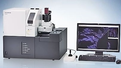
Slide Digitization (SCAN) Learn More
The Olympus VS120 slide loader system is comprised of an Olympus BX-61 motorized system microscope with 2x, 10x, 20x, 40x and 60x oil immersion objective lenses, as well as an Olympus L100-W automatic system slide loader for batch scanning up to 100 slides.
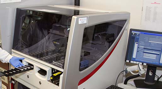
Histology (STAIN) Leica
The BOND RX automates tests including Immunohistochemistry (IHC), In Situ Hybridization (ISH), Immunofluorescence (IF), Fluorescence In-Situ Hybridization (FISH), multiplexing and more!
Tissue Analysis (HALO) Learn More
The HALO® Image Analysis Platform, by Indica Labs, enables quantitative tissue analysis by using digitized tissues, such as those produced by the Olympus VS120 system. HALO® analysis modules enable cellular detection, protein and mRNA quantification and even H-Score calculation.